.png?width=600&name=Ethos%20Health%20Group,%20(1).png "Ethos Health Group")

At our office we recognize the importance of diagnostic imaging in personal injury cases.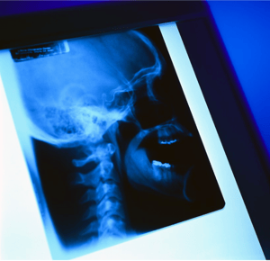
Without demonstrative evidence of injury on x-ray, MRI, CT or other imaging study, it becomes very difficult to establish a true physical link to your client’s reported symptoms.
One of the most challenging tasks in the evaluation and treatment of auto accident injuries has been establish rock solid proof of injury.
You're familiar with the cases I'm talking about: the so-called "soft tissue injuries”, often suffered in relatively low impact collisions with minor damage to the vehicles involved.
How many of these clients have you had come to your office are still in pain weeks or even months after an accident, and their doctors can’t seem to find out why?
An MRI reveals no disc injury, no fractures or dislocations were found on x-ray, but the injured person is still complaining of severe, almost unrelenting pain.
Recent research breakthroughs are providing answers...
 Ligament damage is one of the most common injuries suffered following motor vehicle collisions. Ligaments are injured by sudden loads, and can be damaged in relatively low impact collisions.
Research shows that the forces sustained in crashes as low as 5.7 mph were capable of causing failure of the cervical ligaments.
Ligament rupture results in biomechanics instability and often causes chronic pain. This is precisely the reason that many patients present with no significant findings on MRI or normal x rays, but still complain of severe pain or neurologic symptoms.
Radiographic Evidence of Ligament Damage
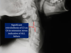
Although a standard x-ray series will not typically show ligament damage, stress view x rays of the cervical or lumbar spine often uncover these so-called “hidden injuries”.
In the cervical spine radiographs are taken with the neck in flexion, extension, and lateral bending to the left and right. These views must be taken on patients who have sustained trauma of any nature in order to evaluate potential ligament failure.
The cervical film at the top of the page is an extension view taken a few days after the patient was involved in a motor vehicle collision. No disc pathology was found on a standard MRI, but these x-rays reveal clear evidence of significant damage to the Anterior Longitudinal Ligament (ALL).
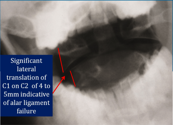
The above x ray is taken through the patient's open mouth as he bends his head to the left.
Normally, the C1 and C2 vertebrae should remain aligned with one another, even as the head is tilted.
This is because the alar and accessory ligaments hold the upper cervical vertebrae in place. These ligaments protect the delicate structures of the spinal cord and surrounding tissue.
As you can see in the above film, there is 4-5 mm of lateral translation of C1 on C2. This represents a severe injury to the upper cervical ligaments, and will be permanent in nature.
This patient will likely require surgical intervention to fuse C1 and C2, and is at significant risk of serious injury should he get in another auto accident, even one at a low speed.
Damage to the Spinal Ligaments Results in Permanent Impairment
Unlike muscle tissue, ligaments have very little blood supply, and therefore are often impossible to heal without surgical intervention.
This fact is so well documented that ligament injury resulting in a loss of motion segment integrity is a rate-able condition according to the AMA Guides to the Evaluation of Permanent Impairment.
In my practice I can't even begin to count the number of patients who have presented with chronic neck pain and degenerative changes as a result of car accidents they experienced 20, 30, even 40 years prior.
Building an Airtight Case with Imaging of Damaged Ligaments
Stress view x-rays are a powerful piece of evidence to show permanent damage that can be linked to a traumatic event.
However, providing compelling diagnostic evidence of ligament damage in the spine involves a three step process.
We have discussed the first, stress view x rays, in detail in this month's newsletter.
In the next two issues of the Whiplash Report we'll discuss Digital Motion X-Ray and Upright MRI, the two tests that allow never before seen proof of traumatic ligament damage in the spine.
In your office I can guarantee you have stacks of files that have not had the proper diagnostic studies to explain your clients symptoms.
Without an expert to properly evaluate and diagnose these previously "hidden injuries" you'll continue to see the same lowball settlement offers the insurance carriers have been offering for years.
Please contact our office to learn more about how we can be an asset to your clients!