.png?width=600&name=Ethos%20Health%20Group,%20(1).png "Ethos Health Group")

 In our last post we touched on a subject that is near and dear to the heart of most personal injury attorneys: the so-called "soft tissue injuries”.
These types of injuries are found in a significant portion of claimants who have been involved in a motor vehicle collision, and are often found in the absence of more objective findings such as fractures, dislocations, or disc herniations.
Most attorneys have stacks of files on their desk that represent clients who continue to report significant pain following crashes, but without any apparent objective proof of injury.
As we discussed, there is exciting new information that is emerging in the area of spinal diagnostics that will quite literally change the way these injuries are viewed forever.
Last month we covered the first step of the three imaging steps to definitively identifying damage to the spinal ligaments. This involved the use of stress view x rays such as flexion, extension, and lateral bending.
The next step involves the use of an imaging study called Digital Motion X Ray, or DMX that gives unprecedented insight into the biomechanics of the spine. DMX studies allow us to pinpoint the exact location and extent of damage to spinal ligaments.
 While DMX is fairly new to most attorneys, it is a technology that has based on technology that has existed since the late 1890s called cine-radiography. Many of you are familiar with a similar technology called. Videoflouroscopy which is used in hospitals and pain management clinics to guide the placement of surgical instruments during procedures like heart catheterizations and epidurals.
DMX has been developed purely as a diagnostic tool, and uses imaging protocols that create a bright white background against which the bones are easily visible.
This type of imaging has been used in trial all over the country, and will pass all Daubert-Frye challenges of admissibility fairly easily. DMX is even used at Walter Reed Army Hospital, one of the premier facilities in the United States where the President receives his medical care.
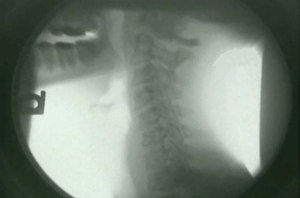
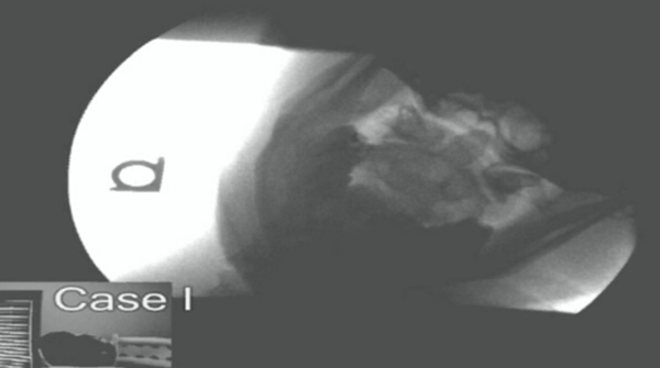
When you watch a DMX study, you are seeing an image of the patient in the lower left hand corner. You’ll see them move their head up and down, left and right, and side to side.
As the patient moves, DMX allows us to clearly visualize the movement of the bones in the spine. In patients who have suffered damage to the spinal ligaments, abnormal motion is present.
You will often see one vertebrae slide several millimeters in relation to another, indicating tearing of the anterior or posterior longitudinal ligaments.
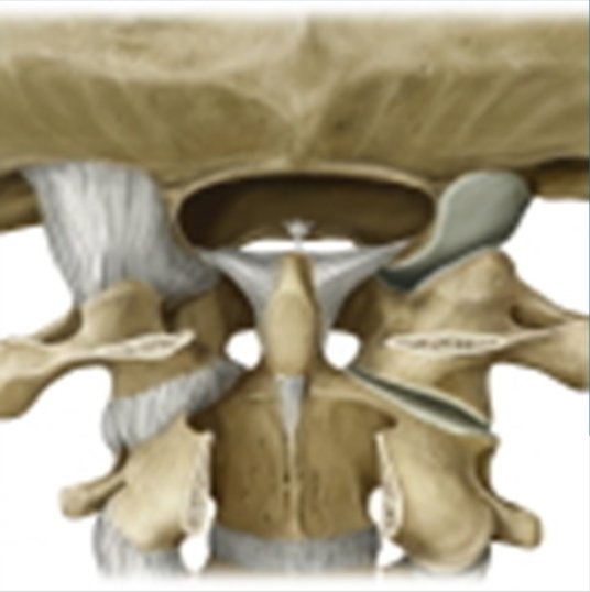
We will often find that the intervertebral foramen (the holes in the spine where the nerves exit) close down in flexion or extension be- cause the capsular ligaments that hold them open have been torn.
The Most Severe Spinal Injuries You’ve Probably Never Heard Of
Verdicts Obtained With The Use of DMX
As I mentioned before, the track record of DMX in the court room is quite impressive. You are no longer limited to a patient saying she is in pain and a treating physician testifying that he believes the patient was indeed injured in a motor vehicle collision. There is now objective evidence that is very visually compelling to a jury that shows the damage.
There are numerous examples of verdicts ranging from $124,500 to $850,000 on cases with quote “normal MRIs" but severe damage to the spinal ligaments shown with DMX.
I hope this topic has piqued your interest and shed some new insight into the management of “soft tissue" cases.
Next month we will cover what I consider to be the nail in the coffin to defense attorneys and adjustors who claim soft tissue injuries should heal in a relatively short period of time and there is no real proof of injury.
The home run on a lot of these cases is a very special type of MRI that a treating physician must be intimately familiar with. This gold standard of spinal imaging will be the final piece of evidence you need to help your injured clients obtain fair compensation for their injuries.
Some of the most severe injuries to the cervical spine are some of the most overlooked by physicians and attorneys alike. Aaron Deshaw DC, JD has lectured extensively on upper cervical ligament injures post MVC.
The C1 and C2 vertebrae contain numerous ligaments such as the alar and transverse ligaments that are easily damaged or torn during car crashes, even at relatively low speeds.
Damage to these ligaments results in symptoms such as headaches, neck pain, blurred vision, double vision, nausea, difficulty sleeping, and numerous other symptoms.
Sound like any of your "soft tissue injury" clients?
The DMX allows these injuries to be clearly visualized by both an adjustor, defense counsel, and jury alike. It is very obvious to watch one bone literally slide off of another as someone flexes their head side to side.
The healing process of any ligament injury is guarded at best. This accounts for the poor long term prognosis exhibited by whiplash patients who have sustained ligament tears in the spine.
Because of its importance with balance and movement, damage to the upper cervical ligaments can be even more devastating.
Once the ligament damage has been identified with the DMX, appropriate specialist referrals may need to occur. In cases of sever upper cervical instability fusion surgery may even be a consideration.
There are very few neurosurgeons in the United States who perform C1-C2 fusions, so these referrals should be chosen carefully to ensure the best possible second opinion has been solicited.